四氯化碳(CCl4)诱导的小鼠肝纤维化和肝硬化是被广泛接受的研究肝纤维化和肝硬化的实验模型。它在许多方面反映了与毒性损伤相关的人类疾病模式,如α-SMA表达、星状细胞活化和关键基质成分(包括胶原蛋白-1、基质金属蛋白酶及其抑制剂TIMPs)已在该模型的发病机制中得到证实。CCl4诱导在肝脏中引起可重现的和可预测的纤维化反应,使其成为抗纤维化和抗肝硬化治疗的临床前药理学研究和肝纤维化-肝硬化-肝癌变化的病理生理学研究的宝贵基础。
Construction of CCl4-induced liver fibrosis model
Exepriment Animal:C57BL/6, 8 weeks old, male
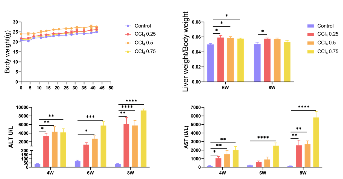
Carbon tetrachloride (CCl4)-induced liver fibrosis model. 8-week-old male C57BL/6 mice were injected intraperitoneally with CCl4 at concentrations of 0.25, 0.5 and 0.75 mL/kg twice a week. Body weight, liver weight/body weight and blood biochemical data of mice after 4, 6 and 8 weeks.
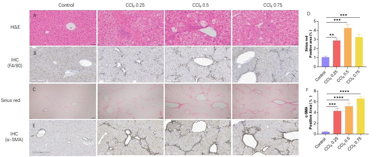
Liver fibrosis model established by CCl4 at 6W . (A) Representative pictures of HE staining showing increased hepatic inflammation after CCl4 induction. Scale bar: 50 um. (B) Representative pictures of Immunohistochemistry showing liver macrophages (kupffer cells) marker F480. (C-D) Representative pictures of sirius red staining showing increased liver fibrosis. Statistic data of sirius red staining. Scale bar: 200 um. (E-F) Representative pictures of Immunohistochemistry showing fibroblast marker α-SMA in liver. Statistic data of α-SMA staining. Scale bar: 300 um.
Reduced liver fibrosis after Obeticholic acid (OCA) treatment
Efficacy Validation on liver fibrosis Mouse Models for OCA (Obeticholic Acid)
(A) Representative pictures of Sirius red staining showing liver fibrosis after 3 weeks of CCl4 induction and OCA treatment. (B) Statistic data of Sirius red staining. Values are expressed as mean ± SEM. *p<0.05.
参考文献
1. Scholten, D., Trebicka, J., Liedtke, C. & Weiskirchen, R. The carbon tetrachloride model in mice. Lab Anim 49, 4-11 (2015).
2. Hernandez-Perez, E., Leon Garcia, P.E., Lopez-Diazguerrero, N.E., Rivera-Cabrera, F. & Del Angel Benitez, E. Liver steatosis and nonalcoholic steatohepatitis: from pathogenesis to therapy. Medwave 16, e6535 (2016).
3. Tsuchida, T., et al. A simple diet- and chemical-induced murine NASH model with rapid progression of steatohepatitis, fibrosis and liver cancer. J Hepatol 69, 385-395 (2018).
4. Farrell, G., et al. Mouse Models of Nonalcoholic Steatohepatitis: Toward Optimization of Their Relevance to Human Nonalcoholic Steatohepatitis. Hepatology 69, 2241-2257 (2019).
Bile duct ligation causes extrahepatic biliary obstruction, which leads to bile duct dilatation and bile stasis. When the pressure in the bile duct increases further, the intrahepatic bile ducts dilate and rupture, the intrahepatic blood vessels are compressed by both the dilated bile ducts and the extravasated bile, the hepatocytes become ischaemic and necrotic, and fibrous tissue proliferates, surrounding the liver lobules and spreading around the hepatocytes, which can eventually lead to cirrhosis.
Construction of the liver fibrosis model by bile duct ligation
Exepriment Animal: C57BL/6, 9 weeks old, male
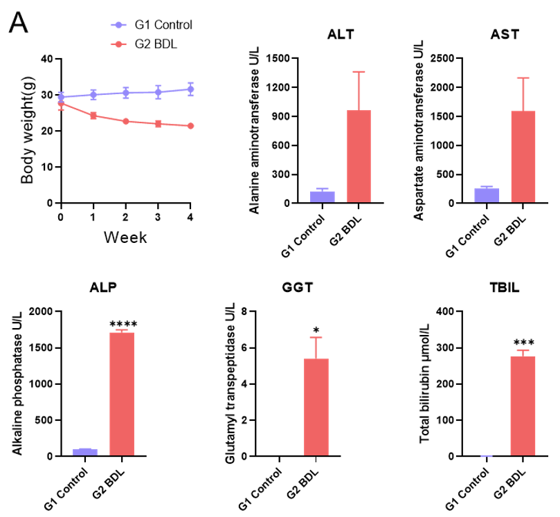
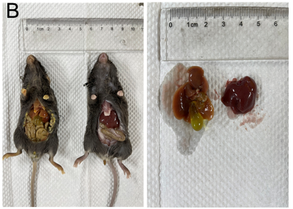
Liver fibrosis model by bile duct ligation (A) ALT, AST, ALP, GGT and TBIL levels in serum.(B)Representative appearance of livers 4 weeks after BDL. Values are expressed as mean ± SEM. *p<0.05, ***p<0.001, ****p<0.0001.
Histologic Assessment of Bile duct ligation induced liver fibrosis model
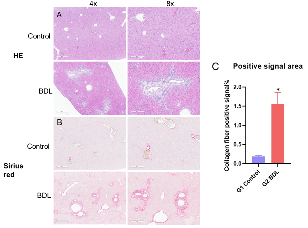
H&E and Sirius red staining after Bile duct ligation 4 weeks (A) Representative pictures of H&E staining. (B) Representative pictures of sirius red staining showing increased liver fibrosis. (C) Positive signal area of collagen fiber. Values are expressed as mean ± SEM. *p<0.05.
Bile duct ligation 4 weeks later (A-B) Representative images of immunohistochemical staining showing F4/80 and positive area. (C-D) Representative images of immunohistochemical staining showing α-SMA and positive area.
Establishment of TAA-induced liver fibrosis model
Thioacetamide (TAA) is a compound widely used to mimic the damage produced in the pathogenesis of liver fibrosis. TAA increases the formation of reactive oxygen species (ROS) through its metabolite thioacetamide sulfur dioxide (TASO2), causing severe oxidative stress as well as lipid peroxidation and the generation of protein carbonyl and DNA adducts. The ROS produced, cause activation of hepatic stellate cells (HSCs), while induces the transdifferentiation of hematopoietic stem cells into myofibroblast-like cells, leading to an imbalance of EMC synthesis and degradation and a continuous fibrotic process.
Construction of the liver fibrosis model by Thioacetamide
Exepriment Animal: C57BL/6, 6-8 weeks old, male
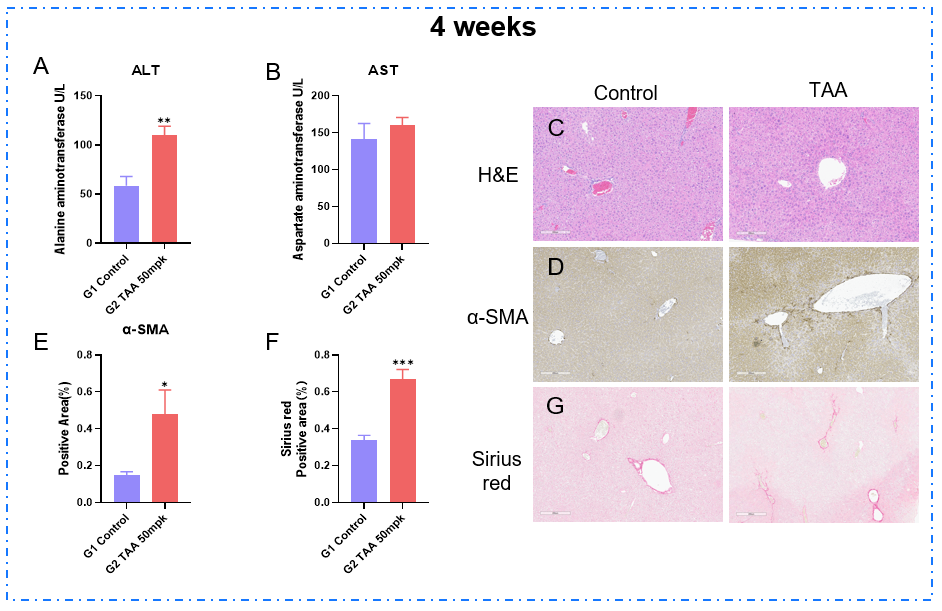
TAA induced liver fibrosis model for 4 weeks. (A-B) ALT and AST levels in serum. (C) Representative pictures of H&E staining. (D-E) Representative images of immunohistochemical staining showing α-SMA and positive area(G-F) Representative pictures of sirius red staining showing increased liver fibrosis and positive area.
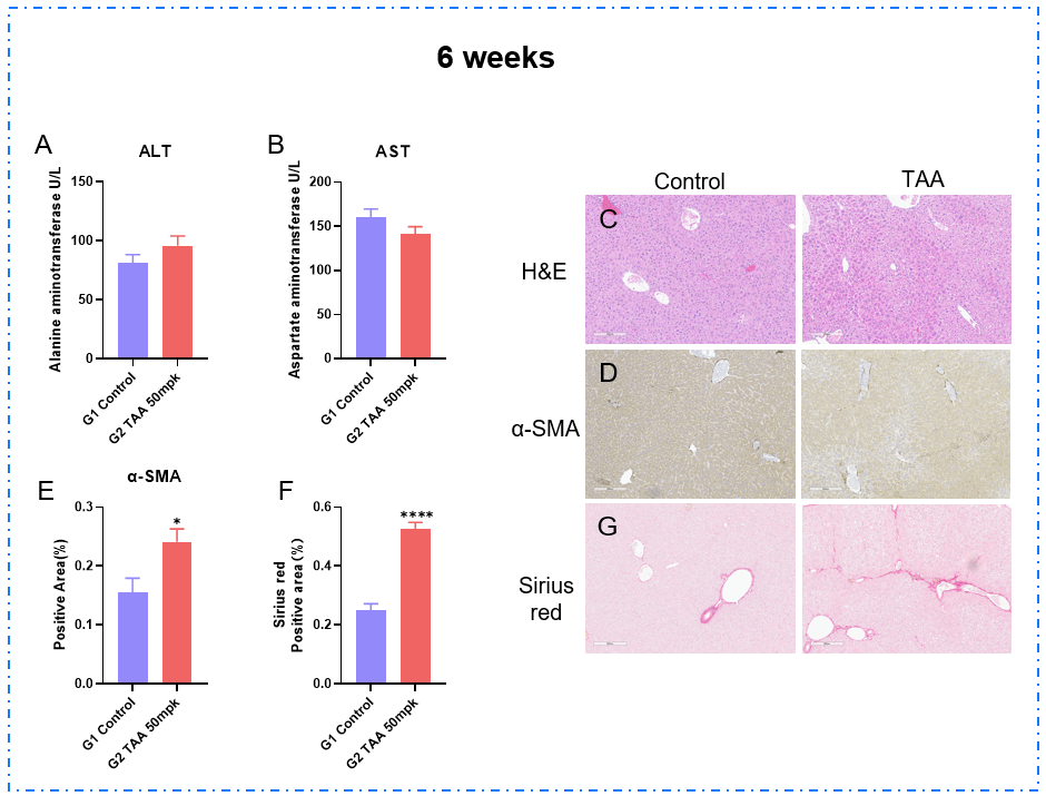
TAA induced liver fibrosis model for 6 weeks. (A-B) ALT and AST levels in serum. (C) Representative pictures of H&E staining. (D-E) Representative images of immunohistochemical staining showing α-SMA and positive area. (F-G) Representative pictures of sirius red staining showing increased liver fibrosis and positive area.